Drugs that can cause Mouth Problems

This search began with a friends question about Zometa and her concern about jaw necrosis with the Biphosphanates. The wider benefit from that search lead to more information related to the wide spread effect that drugs can have on mucosa.
Chemo can cause mouth changes as we know. I came across this list of drugs that can cause mouth changes. I was shocked at some of the drugs on the list. Particularly, b/c I'm on a couple and have had skin issues for awhile. What this list identifies is problems of the mouth. Consider that it is a mucus membrane. More specifically, epithelial tissue. If these drugs have the ability to cause problems for the mouth, they're is a potential for a problem wherever epithelial tissue is found.
The first link is to the list
http://emedicine.medscape.com/article/1080772-overview
The second link is to everything you ever(or never) wanted to know about Epithelial tissue.
http://anatomyandphysiologyi.com/epithelial-tissue/
I'll use me as an example. I have several forms of eczema. All were diagnosed at different times. Eczema is a problem of epithelial tissue. Times that the eczema was worse was related to different periods. One was specific to the only chemo I had which lasted for months. Another was a problem after Radioactive Iodine131 for Thyroid cancer. But until now I never associated that any other drug could be related. Two drugs that are on the list are the benzo's and slow channel calcium blocker amlidopine. The connection here is that a drug, the combination of drugs, the combination of drugs plus chemo & RAI131--all could have been a source of my problem at a given time. For example, the RAI131 stays around along time, during that time I took benzo's and amlidopine. Individually they can cause problems. Three at the same time could explain the many months of hell.
No doc-PCP, dermatologist, cardiologist, MO, ever questioned what drugs I was on as an origin or a contributing factor. They each took care of their body part. Hope this makes sense to you. It's an eye opener for me.
I'm going to put the whole list here for review.
Comments
-
All of the following posts are copied and pasted from the Medscape link.
An estimated 2-4% of hospital admissions are related to adverse drug reactions. Mucocutaneous eruptions are often central to these untoward reactions, and an ever-expanding list of medications is linked to pathologic reactions in the oral and perioral region. These adverse drug reactions have a broad spectrum of clinical manifestations that can mimic those of other disease states, including both local and systemic conditions.[1, 2, 3, 4, 5]
Fortunately, several patterns of disease have been identified, and these can assist the clinician in determining a possible cause-and-effect relationship with a particular medication or group of medications. The clinical patterns of adverse drug reactions of the oral cavity include xerostomia, swelling, nonspecific ulceration, vesiculobullous or ulcerative mucositis that mimics other disease states, nonspecific vesiculoulcerative mucositis, pigmentation, gingival enlargement, and medication-related osteonecrosis of the jaws.
Oral drug-reaction patterns with associated drugs and drug classes
Xerostomia
Agents include antidepressants and antipsychotics, antihypertensives, antihistamines, anticholinergics, and decongestants.
Swelling
Agents include penicillins, aspirin, sulfa drugs, and ACE inhibitors.
Nonspecific ulceration and mucositis
"" frameborder="0" marginwidth="0" marginheight="0" scrolling="no" style="border: 0px currentColor; border-image: none; vertical-align: bottom;">Agents include antineoplastics (methotrexate, 5-fluorouracil, doxorubicin, and melphalan); barbiturates; dapsone; phenazone derivatives; phenolphthalein; sulfonamides; tetracyclines; NSAIDs (indomethacin, salicylates, gold salts, naproxen); meprobamate; methyldopa; penicillamine; phenylbutazone; propranolol; spironolactone; thiazides; tolbutamide; alendronate; captopril; phenytoin; and (by direct contact) compounds containing aspirin, hydrogen peroxide, or phenol.
Vesiculobullous or ulcerative lesions that mimic other immunologic diseases
Reactions may include lichen planus–like, erythema multiforme (EM)–like, pemphigoid-like, pemphigus -like, and lupus erythematosus (LE)–like reactions.
Lichen planus–like[6, 7, 8] agents may include the following:
- Antialcoholism (cyanamide)
- Antibiotics (aminosalicylate sodium, isoniazid, rifampin, streptomycin, tetracyclines)
- Anticonvulsants (carbamazepine, oxacarbazepine, phenytoin, valproate)
- Antidiabetics (chlorpropamide, glipizide, insulin, tolbutamide)
- Antidiarrheals (bismuth)
- Antifungals (amphotericin B, ketoconazole)
- Antihistamines (cimetidine, cinnarizine, triprolidine)
- Antihypertensives (atenolol, captopril, chlorothiazide, enalapril, furosemide, hydrochlorothiazide, methyldopa, metoprolol, oxprenolol, propranolol, spironolactone)
- Antihyperuremics (allopurinol)
- Antimalarials (chloroquine, hydroxychloroquine, pyrimethamine, quinacrine, quinidine)
- Antiretroviral (zidovudine)
- Calcium channel blocker (flunarizine)
- Chemotherapeutics (dactinomycin, imatinib, palladium)
- Diuretic (furosemide)
- Immunomodulators (dapsone, gold salts, interferon alfa, levamisole, penicillamine)
- NSAIDs (acetylsalicylic acid, para-aminosalicylic acid, diflunisal, ibuprofen, indomethacin, naproxen, rofecoxib, sulindac)
- Psychiatrics (benzodiazepines, tricyclic antidepressants, levopromazine, lithium, lorazepam, phenothiazines, pyritinol)
Erythema multiforme (EM)–like agents may include the following:
- Antibiotics (antimalarials, penicillins, sulfonamides, tetracyclines)
- Allopurinol
- Barbiturates
- Protease inhibitors
- NSAIDs
Pemphigoid-like[9] agents may include the following:
- Antirheumatics (penicillamine, ibuprofen, phenacetin)
- Cardiovascular drugs (furosemide, captopril, clonidine)
- Antibiotics (penicillins, sulfonamides)
- Antimicrobials
- Thiol-containing drugs
- Sulfonamide derivatives
Pemphigus -like[10] agents may include the following:
- Alpha-mercaptopropionylglycine
- Ampicillin
- Captopril
- Cephalexin
- Ethambutol
- Glibenclamide
- Gold
- Heroin
- Ibuprofen
- Penicillamine
- Phenobarbital
- Phenylbutazone
- Piroxicam
- Practolol
- Propranolol
- Pyritinol chlorohydrate
- Rifampin
- Theobromine
Lupus erythematosus (LE)–like agents may include the following:
- Carbamazepine
- Chlorpromazine
- Ethosuximide
- Gold
- Griseofulvin
- Hydantoins
- Hydralazine
- Isoniazid
- Lithium
- Methyldopa
- Penicillamine
- Primidone
- Procainamide
- Quinidine
- Reserpine
- Streptomycin
- Thiouracils
- Trimethadione
Pigmentation
Agents include amiodarone; antimalarials (chloroquine, hydrochloroquine, hydroxychloroquine, quinacrine, quinidine); busulfan; clofazimine; cyclophosphamide; estrogen; imatinib; ketoconazole; minocycline; phenolphthalein; chlorpromazine; and zidovudine.
Gingival enlargement
Agents include calcium channel blockers (amlodipine, bepridil, diltiazem, felodipine, isradipine, nicardipine, nifedipine, nimodipine, nisoldipine, nitrendipine, oxidipine, verapamil); other dihydropyridines (bleomycin); cyclosporine; phenytoin; and sodium valproate.
-
Continuing list
Xerostomia, or dry mouth, is the most common adverse drug-related effect in the oral cavity. To date, xerostomia has been associated with more than 500 medications. The synergistic effects of medications have been recognized and are increasingly common in elderly patients taking multiple medications (polypharmacy). In addition, habits such as smoking, alcohol consumption, and even long-term use of caffeinated drinks may contribute to oral dryness or the perception of dryness.
General drug classes that are strongly associated with xerostomia include antidepressants and antipsychotics, antihypertensives, antihistamines, and anticholinergics.
Unfortunately, these associations are confounded by the fact that xerostomia is a subjective complaint, and some patients who report oral dryness may have apparently normal salivary gland function. Other patients whose mouths appear to be clinically dry may have no complaints. In addition, a certain amount of acinar atrophy and decreased glandular secretions are considered a normal part of the aging process. Given these variations, clinical tests to measure salivary flow (at rest and under stimulation) should be used cautiously in assessing a patient with suspected drug-induced xerostomia.
Furthermore, the possibility of an underlying autoimmune etiology (eg, Sjögren syndrome) should also be considered in xerostomic patients, especially those who also present with xerophthalmia (ie, dry eyes) or evidence of parotid swelling. Appropriate laboratory studies or biopsy of the labial minor salivary glands or parotid glands may be helpful in establishing or excluding an autoimmune etiology.
Importantly, patients with xerostomia are also at increased risk for oral candidiasis, a superficial infection that may cause mucosal sensitivity or discomfort, as well as tooth decay that usually affects the cervical (gumline) and root surfaces. Cultures and oral cytologic tests can be useful in assessing possible cases of xerostomia-associated candidiasis. Meticulous oral hygiene, regular dental checkups, and the use of topical fluoride rinses are recommended to reduce the risk of dental decay.
-
Swelling
Several drugs can induce type I hypersensitivity reactions, or disease mediated by immunoglobulin E mast cells, that can range from isolated swelling of the oral tissues to full-blown anaphylaxis. Around the mouth, the lips are the most frequently involved site, followed by the tongue. Among the most common offending agents are ACE inhibitors, penicillin and penicillin derivatives, cephalosporins, barbiturates, and aspirin and other NSAIDs. Affected mucosa typically appears edematous and erythematous within minutes or hours after exposure to the offending drug. Similar contact reactions to latex had become increasingly problematic in oral health care settings until the recent shift towards non-latex replacement materials such as vinyl or nitrile rubber.
ACE inhibitors are also associated with oral swelling of a nonallergic nature. This reaction, which is possibly secondary to alterations in local bradykinin levels or C1 esterase inhibitor function, is reported in less than 1% of patients, with an increased risk noted among blacks in the United States. Although swelling is most commonly seen in the first weeks of therapy, it may occur within hours of the initial exposure or after an extended period of drug use. As with the type I reactions, the lips are the most typical site of involvement. The tongue and oropharyngeal tissues can also be affected, with a potentially fatal outcome.
-
Nonspecific Ulceration and Mucositis
Epithelial necrosis and ulceration may result from direct application of over-the-counter medications such as aspirin, hydrogen peroxide, potassium tablets, and phenol-containing compounds to the mucosa. Aspirin is often used by patients seeking relief from dental pain. The affected mucosa appears whitish and corrugated, with erosion and ulceration of the more severely damaged areas. The associated discomfort can be severe enough to require treatment with a strong analgesic and/or a local anesthetic.
Fixed drug eruptions in the oral cavity often initially appear as areas of edema and erythema that lead to localized, nonspecific ulceration. The labial mucosa is most commonly involved, and a clinical course of recurrence at the same site after drug use is diagnostically helpful, but this relationship is not always easy to establish. Extraoral involvement, including involvement of the cutaneous surface and other mucosal surfaces, may occur.
A number of drugs are implicated in the development of nonspecific ulceration and oral mucositis, and the lesions are often associated with an equally nonspecific histologic appearance at biopsy. These include barbiturates, beta-blockers, dapsone, NSAIDs, phenazone derivatives, thiazide derivatives, phenolphthalein, sulfonamides, and tetracyclines. Recently, oral mucositis affecting the buccal mucosa and palate was reported in association with use of tadalafil.[11] Ulceration of the oral mucosa is a common adverse effect of a wide variety of antineoplastic agents, including methotrexate, 5-fluorouracil, doxorubicin, and melphalan and may lead to discontinuation of the chemotherapeutic agent.[12]
-
Vesiculobullous or Ulcerative Lesions That Mimic Other Immunologic Diseases
Oral drug reactions that bear striking clinical, histopathologic, and even immunopathologic resemblance to idiopathic lichen planus, erythema multiforme (EM), pemphigoid, pemphigus, and lupus erythematosus (LE) are well recognized, and the list of reactions in each category is constantly expanding. Clinically, any oral site can be affected; however, the posterior buccal mucosa (cheeks), the lateral borders of the tongue, and the alveolar mucosa are most commonly involved. Lesions may be isolated, although bilateral and occasionally symmetric involvement is not uncommon.
Lichen planus – like (lichenoid) reactions
Initially described in association with antimalarial medications, lichen planus–like or lichenoid drug reactions have subsequently been reported in association with many other agents. Both papuloreticular and erosive manifestations may be observed; the latter is characterized by shallow irregular ulcerations or erosions with a peripheral border of fine keratotic striae that often appear to radiate from the center of the lesion. Although drug-induced lichenoid reactions tend to be erosive and unilateral compared with the typical bilateral presentation in idiopathic lichen planus, these associations are not consistently observed.
Currently, NSAIDs and ACE inhibitors appear to be among the most frequently cited offenders (see Oral drug-reaction patterns and associated drugs and drug classes in Introduction). Interestingly, agents used in the treatment of lichen planus (eg, hydroxychloroquine, dapsone, levamisole) have themselves led to adverse lichenoid eruptions.
Erythema multiforme (EM) – like reactions
Drug-induced EM represents approximately 25% of all reported cases. As with idiopathic or virally induced cases (the latter often due to the herpes simplex virus), the disease has a rapid onset with a variable expression that can range from lesions limited to the oral mucosa to widespread mucocutaneous involvement. Drug-induced EM is frequently linked to agents such as sulfonamides, sulfonylureas, and barbiturates, among others (see Oral drug-reaction patterns and associated drugs and drug classes in Introduction).
Oral lesions start as erythematous macules or patches that lead to short-lived vesicles or bullae, followed by ragged and shallow ulcerations that may become extensive. Hemorrhagic ulceration and crusting of the labial vermilion zone is common. Any intraoral site can be involved, but the attached gingival and palatal tissues are often relatively spared. Lesional discomfort can lead to decreased fluid intake and dehydration. A variety of lesions, including classic target or bull's eye lesions, can affect the skin.
More severe forms of disease include EM major (Stevens-Johnson syndrome) and toxic epidermal necrolysis (TEN, Lyell syndrome). In addition to the oral and skin lesions, conjunctivitis of the ocular mucosa and urethritis of the genital mucosa are typical findings in patients with EM major. Over three-quarters of EM major cases and virtually all cases of TEN represent adverse drug reactions. In TEN, widespread mucocutaneous epidermolysis results in diffuse bullae formation and subsequent denudation that affects significant proportions of the skin and mucosal surfaces. Severe dehydration, electrolyte imbalances, and secondary infections are potentially life-threatening sequelae, and even with aggressive therapy the mortality rate is greater than 30%.
Pemphigoid-like reactions
Pemphigoid-like reactions can be limited to the oral mucosa, or they can affect other mucosal or cutaneous sites. Clinically, lesions appear as relatively sturdy vesicles or bullae that break down into shallow ulcerations. Generalized or multifocal involvement of the gingival tissues may be observed, with marked erythema and erosion of the superficial gingiva, a pattern that has been called desquamative gingivitis. Thiol-containing drugs and sulfonamide derivatives are among the most commonly involved medications, as are the therapeutic classes of NSAIDs, cardiovascular agents, antimicrobials, and antirheumatics (see Oral drug reaction patterns with associated drugs and drug classes in Introduction).
Circulating autoantibodies to basement membrane components may or may not be detectable. The relatively young age of the patients at onset (usually less than 70 years) may help distinguish this disease from idiopathic bullous pemphigoid or idiopathic cicatricial pemphigoid, because these latter conditions typically appear in persons aged 60-80 years. In addition, oral involvement may be more common in drug-induced bullous pemphigoid than in the idiopathic counterpart.
Pemphigus-like reactions
Pemphigusike reactions can have features of either pemphigus vulgaris or pemphigus foliaceous, although pemphigus foliaceous is uncommon in the oral cavity. Thiol-containing drugs are the most common cause of pemphiguslike reactions (see Oral drug-reaction patterns and associated drugs and drug classes in Introduction). In drug-induced pemphigus vulgaris, the relatively fragile vesicles are rarely observed at clinical examination, and most cases are characterized by irregular ulcerations with ragged borders that may coalesce to involve large areas of the mucosa. Patients may have circulating autoantibodies to the desmosomal components.
Lupus erythematosus (LE) – like reactions
Drug-induced LE is a well-recognized adverse reaction that is most commonly associated with procainamide and hydralazine, although more than 70 medications are implicated (see Oral drug-reaction patterns and associated drugs and drug classes in Introduction). Clinically, the oral lesions of drug-induced LE may simulate those of erosive lichen planus, with irregular areas of erythema or ulceration bordered by radiating keratotic striae. These lesions may affect the palate, buccal mucosa, and gingival or alveolar tissues. The rarity of lichen planus on the hard palate may be helpful in differentiating it from drug-induced LE.
-
Pigmentation
Discoloration of the oral mucosa after drug use may be due to direct melanocytic stimulation, the deposition of pigmented drug metabolites, or both. This reaction has long been recognized with antimalarial agents, particularly chloroquine, hydroxychloroquine, quinacrine, and quinidine (see Oral drug-reaction patterns and associated drugs and drug classes in Introduction). Typically, such pigmentation is most notable on the midposterior regions of the hard palate, appears bluish-black to brown, and may be bilateral. A similar pattern is described with tranquilizers, especially chlorpromazine, as well as in patients with chronic myeloid leukemia who have been treated with imatinib.[13]
Macular or diffuse oral pigmentation may occur after the treatment of HIV disease with zidovudine, clofazimine, and ketoconazole. Minocycline use may be associated with bluish-gray to brownish mucosal pigmentation. Staining of underlying bony structures and tooth roots and enamel can occur. Staining of the bone and tooth roots is believed to play a major role in the most notable sites of discoloration, such as the alveolar, gingival, and palatal tissues. Yet, staining of the mucosa alone has been reported in sites such as the tongue.
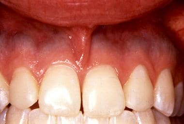 Minocycline-associated pigmentation in a patient who had used the drug for several months to treat severe acne. Note the bluish gray hue of the alveolar mucosa superior to the attached (pink) gingival mucosa. Courtesy of Jack Caton, DDS, Rochester, NY. Previous
 -
Gingival Enlargement
Diffuse, non-neoplastic enlargement or overgrowth of the gingival tissues was initially recognized in patients who were using phenytoin. More recently, calcium channel blockers (members of the dihydropyridine class of medications), cyclosporine, and the antiepileptic drug sodium valproate have been associated with this reaction. Within the calcium channel blocker family, nifedipine, diltiazem, verapamil, and amlodipine are among the most commonly reported causative agents (see Oral drug-reaction patterns and associated drugs and drug classes in Introduction).
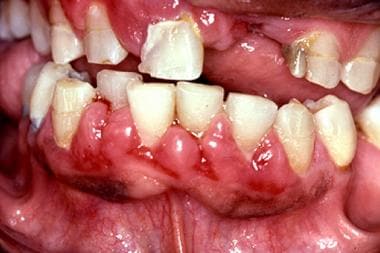 Gingival enlargement in a 41-year-old man with a several-year history of using calcium channel blockers. Courtesy of Carl Allen, DDS, Columbus, Ohio.
 Gingival enlargement is not observed in all patients. The prevalence is 25-50%, and no clear relationship has been established between the dose of the drug and the severity of the overgrowth. Synergistic effects have been reported with the use of 2 or more suspected agents. Tissue enlargement typically occurs by 1-3 months after drug therapy is initiated and begins in the superficial gum tissues between the teeth (interdental papillae). Anterior segments are more frequently involved than posterior areas, but generalized involvement is not uncommon.
An inverse relationship exists between oral hygiene and the degree of gingival enlargement associated with these drugs. Although good oral hygiene typically does not prevent enlargement in susceptible individuals, it can often limit the severity of the response to acceptable levels. Although cessation or substitution of the drug may lead to regression, surgical removal of the excess tissue (ie, gingivectomy) may be necessary to permit adequate oral hygiene in certain individuals.
-
Medication-Related Osteonecrosis of the Jaws
Osteonecrosis of the jaws (ONJ) is a recognized adverse effect of bisphosphonate (BP) therapy but more recently has been described in association with other antiresorptive, antiangiogenic, and immunomodulatory medications.[14, 15, 16, 17, 18] Clinically, ONJ is characterized by prolonged exposure and necrosis of portions of the jawbone(s). BPs significantly reduce the rate of bone turnover, primarily by inhibiting osteoclastic activity. Given the relatively high metabolic demands of the jaws, it is thought that sustained use of BP drugs may suppress the ability of the jawbones to fulfill normal maintenance and repair functions, especially in the presence of the thick, complex microflora found in the oral cavity.
BP drugs are widely used, most commonly in the treatment of osteoporosis and other metabolic bone diseases. The greatest risk for ONJ, however, appears to be among cancer patients. A number of malignant neoplasms, such as multiple myeloma, carcinoma of the breast, and carcinoma of the prostate, have a recognized propensity for skeletal involvement. BP therapy significantly reduces local and metastatic spread of these skeletal lesions, as well as associated morbidity and mortality. Cancer patients are treated with intravenous forms of BP and at much higher doses than those used for metabolic bone conditions. These factors may help explain why more than 90% of published cases of ONJ have occurred in the setting of cancer therapy.
ONJ has also been associated with denosumab, a monoclonal antibody that inhibits RANK ligand-stimulation of osteoclast function and viability. Similar to BP drugs, more cases of denosumab-related ONJ have been reported in patients with cancer than in those with osteoporosis.
ONJ has also been reported in cancer patients taking antiangiogenic agents, including bevacizumab, sunititib, and sorafenib, and the combined use of both antiresorptive and antiangiogenic drugs may increase the risk for bone exposure and necrosis.[19]
See the image below.
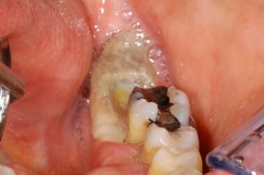 Exposure of the posterior left mandible in a 51-year-old woman with a history of breast cancer and treatment with intravenous bisphosphonate (zoledronic acid [Zometa]).
 Medication-related ONJ is characterized by exposure and devitalization of bone in the mandible or maxilla that may be asymptomatic or associated with significant pain. The mandible is involved more commonly than the maxilla (2:1); however, multifocal presentations involving both jaws or multiple sites in a single jaw are not uncommon. Prior dental surgery or extraction is reported in 60% of patients, with the remaining cases arising spontaneously. Clinically, the lesions appear as areas of exposed, yellow-white bone with margins that vary from smooth to ragged. Secondary infection of lesional sites may develop, with the development of intraoral sinus tracts or even cutaneous fistulae.
Information regarding treatment and follow-up of medication-related ONJ remains limited. Conservative measures such as gentle debridement of superficial necrotic bone, topical antibiotic rinses, and systemic antibiotic therapy to control areas of active infection have shown the greatest success. While early studies found little success with aggressive surgical therapy or adjunctive measures such as hyperbaric oxygen therapy, more recent evidence suggests that they may be helpful in selected cases when combined with primary mucosal wound closure and local and systemic antibiotics.
In order to prevent or reduce the risk for medication-related ONJ, all cancer patients scheduled to begin intravenous antiresorptive therapy should receive a careful dental examination to identify and eliminate active or potential sources of infection. This approach reduces the need for subsequent extractions or dentoalveolar surgery, which are known risk factors for the development of ONJ. During antiresorptive or anticancer treatment, all patients should receive periodic intraoral examinations to detect the earliest evidence of ONJ. When ONJ is identified, conservative treatment measures that minimize bone manipulation or exposure are generally favored over aggressive surgical intervention, unless the latter is warranted on the basis of pain, extensive bone exposure, presence of active infection or purulence, or combinations of these factors.
-
Bump
-
bump
-
Thank you for pulling this information up. No one said a word to me about my teeth on;ly the dentist. I do have dry mouth and use a special tooth past for it. My mouth looks horrid, I anxiously await having a normal smile again.
SAS thanks for all that you do.
Ginger
-
") ginger
ginger -
Bump
-
Bump
-
Bump
-
Thank you for the information above. I am wondering if other women can chime in and describe their own dental procedures and tooth/jaw pain, and what they think MIGHT have caused it. I am desperate as I am in extreme pain. Is this a s/e of the CMF chemo or some part of treatment? I have always had decent teeth, but now, after 3 years of active treatment (I'm Stage IV) I have "condensed osteoporosis in my right lower mandible and have suddenly developed pain under a tooth that I have had a root canal & crown on for years. It is giving me extreme pain, that my narcotics are not helping.
My Oncologist doesn't want to do anything about it, because he's worried about Necrotic Jaw Syndrome (NJS) that 1% of women on Denosumab get. And my low WBC and risk for infection with dental procedures. My dentist won't touch me without the Oncologists permission. I want the tooth pulled, but I'm probably going to have to go against my Oncologists advice and do it anyways.
Does anyone have any similar experience with this or NJS? I'm at my wits end. I use Orajel and Anebosol and salt water swish and spits.Thanks in advance for and advice or related stories.
-
Hi AKShelley,
I've often wondered how you're doing and am sad to hear that you too are facing terrible dental pain. I'm not but one of a close friend has all year long - it's as devastating to her as progressing abdominal and bone mets. Like you, she has ONJ or as you call it in Alaska, NJS.
I wrote about it at the Ibrance thread just a month ago:
I've become a warrior to speak up about ONJ (osteonecrosis of the jaw) in those with bone mets who take the bone drugs Zometa (or other bisphosphonates) or Xgeva.
Shelley, I know you want to act on your own behalf and hope you'll find a dental oncologist to perform this delicate work. The damage often extends beyond the tooth and you really don't want to end up with jaw necrosis.
Two close friends with MBC have both endured torture, because of missed diagnosis and mistreatment of ONJ.
They and I are now encouraging everyone with bone mets and dental problems requiring major dental work, including tooth extraction, to see dental oncologists, if available in your region. Most major cancer centers seem to have them or be able to refer.
Please, don't get dental work done by any professional who's not skilled in the specific needs of those who've engaged these strong bone drugs.
Take your time to find proper medical care for dental problems when at risk for ONJ.
End of lecture, no end of loving kindness and high regard, Stephanie
Shelley, I know you want to act quickly on your own behalf and hope you'll find a dental oncologist to perform this delicate work. The damage often extends beyond the tooth and you really don't want to end up with jaw necrosis.
Sending best wishes and loving kindness this early morning.
I'm up with nausea and a sore, dry mouth - xeorstomia from frequent procedures.
Your friend, Stephanie
-
Bump
-
Thanks for the bump. I have eczema as well, and didn't see the connection to the inside of my mouth. I was not aware of so many drugs interfering with a healthy mouth. I've had some issues just last week with mouth sores and was puzzled about their origin. This information has been helpful.
-
Tarheel
") As stated from the first post it's totally taken from another site. Let us know if something in particular helps sassy
As stated from the first post it's totally taken from another site. Let us know if something in particular helps sassy -
Bump
-
Akshelly Im in terrible pain too. I had to get deep cleaning done before I started xgeva so I did that a few weeks ago. I started ibrance xgeva and was already on faslodex. I took 18 pills out if 21 then I had to call onc and tell him i was in too much pain si we stopped the ibrance until my next cycle and he said we would skip the xgeva injection. I called the dentist thursday and he saw me and said i had sime ulcers. I dont understand how ulcers can cause jaw and teeth pain. My haw is swollen to. I was rinsing with chlorahexidine and winder if that was causing the problem. My inc said he hasnt heard of that side effect on ibrance but im pretty sure its a listed side effect. I hope you feel better. I have been rinsing with baking soda and water and salt and water. Ive been eating 4 ibuprophen every 8 hours and taking a pain pill when im not at work. Ive also put ice packs on my jaw. I think im starting to get better but not sure yet. I still cant eat. Thank Sas for bumping this and for all of the info.
-
Your welcome Mel. Please, consider bumping this once and awhile if I haven't
-
-
Transferred post written by LoverofLife. Esophageal reflux/GERDS can impact the mouth and the posterior throat
Sep 29, 2017 10:55PM Loveroflife wrote:
Oooh the light bulb just came on. May I add my thoughts....,some drugs can actually cause reflux ( calcium channel blockers, potassium supplements for example).
From Mayo:


-
Bump
-
Bump
-
I had 6 months of Herceptin, Progetta and Taxotere and lost my taste right away. Food tastes terrible and even though I have finished my triple chemo drugs I still hate eating because of the horrible taste food leaves in my mouth. I am only on Herceptin every 3 weeks now but it isn't supposed to give me any side affects so my taste is supposed to come back. So far that isn't happening yet and I am so frustrated. Did anybody else have this problem and if so how long did it take before your mouth went back to normal?
-
Akbornlady-
Welcome to BCO! That sounds incredibly frustrating, we're sorry that you're still dealing with this. Hopefully another member who's been through it has some advice/words of encouragement about how long it'll take for your mouth to return to normal.
The Mods
-
I've added four sources. Taste and smell are problems without an easy answer. I so get your problem b/c I have an alteration of both. Mine are from Chronic sinusitis, smoking, environmental & food allergies. My newest interfering problem with smell and taste, is sleep apnea with CPAP.
Consider yourself a detective at this point. You identify the culprit of your problem as being related to chemo. The importance of doing a complete detective job is to make sure you have uncovered all the clues. Yes, you attribute the loss to chemo, but they're may now be extenuating circumstances. i.e. other drugs, gum disease caused by chemo/radiation. Reading a wide variety of info is important to uncover all the clues.
The first article, is related to that specific problem of chemo. The second article, is with Carl Philpott. I like this article, because he has been researching the problem for a very long time. He actually has a clinic. His question and answer session in the interview is very enlightening.
Taste Changes During Chemotherapy
What to do when food loses its flavor during chemotherapy
By Lisa Fayed | Reviewed by Doru Paul, MDUpdated March 20, 2017
https://www.verywell.com/taste-changes-during-chemotherapy-513895
/////////////////////////////////////////////////////////////////////////////////////////////////////////////////
This article as sent me on a quest to find other Olfactory Taste clinics. Carl Philpotts' clinic is in the UK
The Unappreciated Senses
The world is a bleak place if you lose your sense of taste or smell.
By Mick O'Hare Interview with Carl Philpott
May 12, 2013


Categories
- All Categories
- 679 Advocacy and Fund-Raising
- 289 Advocacy
- 68 I've Donated to Breastcancer.org in honor of....
- Test
- 322 Walks, Runs and Fundraising Events for Breastcancer.org
- 5.6K Community Connections
- 282 Middle Age 40-60(ish) Years Old With Breast Cancer
- 53 Australians and New Zealanders Affected by Breast Cancer
- 208 Black Women or Men With Breast Cancer
- 684 Canadians Affected by Breast Cancer
- 1.5K Caring for Someone with Breast cancer
- 455 Caring for Someone with Stage IV or Mets
- 260 High Risk of Recurrence or Second Breast Cancer
- 22 International, Non-English Speakers With Breast Cancer
- 16 Latinas/Hispanics With Breast Cancer
- 189 LGBTQA+ With Breast Cancer
- 152 May Their Memory Live On
- 85 Member Matchup & Virtual Support Meetups
- 375 Members by Location
- 291 Older Than 60 Years Old With Breast Cancer
- 177 Singles With Breast Cancer
- 869 Young With Breast Cancer
- 50.4K Connecting With Others Who Have a Similar Diagnosis
- 204 Breast Cancer with Another Diagnosis or Comorbidity
- 4K DCIS (Ductal Carcinoma In Situ)
- 79 DCIS plus HER2-positive Microinvasion
- 529 Genetic Testing
- 2.2K HER2+ (Positive) Breast Cancer
- 1.5K IBC (Inflammatory Breast Cancer)
- 3.4K IDC (Invasive Ductal Carcinoma)
- 1.5K ILC (Invasive Lobular Carcinoma)
- 999 Just Diagnosed With a Recurrence or Metastasis
- 652 LCIS (Lobular Carcinoma In Situ)
- 193 Less Common Types of Breast Cancer
- 252 Male Breast Cancer
- 86 Mixed Type Breast Cancer
- 3.1K Not Diagnosed With a Recurrence or Metastases but Concerned
- 189 Palliative Therapy/Hospice Care
- 488 Second or Third Breast Cancer
- 1.2K Stage I Breast Cancer
- 313 Stage II Breast Cancer
- 3.8K Stage III Breast Cancer
- 2.5K Triple-Negative Breast Cancer
- 13.1K Day-to-Day Matters
- 132 All things COVID-19 or coronavirus
- 87 BCO Free-Cycle: Give or Trade Items Related to Breast Cancer
- 5.9K Clinical Trials, Research News, Podcasts, and Study Results
- 86 Coping with Holidays, Special Days and Anniversaries
- 828 Employment, Insurance, and Other Financial Issues
- 101 Family and Family Planning Matters
- Family Issues for Those Who Have Breast Cancer
- 26 Furry friends
- 1.8K Humor and Games
- 1.6K Mental Health: Because Cancer Doesn't Just Affect Your Breasts
- 706 Recipe Swap for Healthy Living
- 704 Recommend Your Resources
- 171 Sex & Relationship Matters
- 9 The Political Corner
- 874 Working on Your Fitness
- 4.5K Moving On & Finding Inspiration After Breast Cancer
- 394 Bonded by Breast Cancer
- 3.1K Life After Breast Cancer
- 806 Prayers and Spiritual Support
- 285 Who or What Inspires You?
- 28.7K Not Diagnosed But Concerned
- 1K Benign Breast Conditions
- 2.3K High Risk for Breast Cancer
- 18K Not Diagnosed But Worried
- 7.4K Waiting for Test Results
- 603 Site News and Announcements
- 560 Comments, Suggestions, Feature Requests
- 39 Mod Announcements, Breastcancer.org News, Blog Entries, Podcasts
- 4 Survey, Interview and Participant Requests: Need your Help!
- 61.9K Tests, Treatments & Side Effects
- 586 Alternative Medicine
- 255 Bone Health and Bone Loss
- 11.4K Breast Reconstruction
- 7.9K Chemotherapy - Before, During, and After
- 2.7K Complementary and Holistic Medicine and Treatment
- 775 Diagnosed and Waiting for Test Results
- 7.8K Hormonal Therapy - Before, During, and After
- 50 Immunotherapy - Before, During, and After
- 7.4K Just Diagnosed
- 1.4K Living Without Reconstruction After a Mastectomy
- 5.2K Lymphedema
- 3.6K Managing Side Effects of Breast Cancer and Its Treatment
- 591 Pain
- 3.9K Radiation Therapy - Before, During, and After
- 8.4K Surgery - Before, During, and After
- 109 Welcome to Breastcancer.org
- 98 Acknowledging and honoring our Community
- 11 Info & Resources for New Patients & Members From the Team